
Common Terms
- Ligament: a ligament is a strong tissue that connects a bone to another bone, creating stability for a joint.
- Tendon: a tendon is the thick tissue that connects the belly of a muscle to a bone
- Cartilage: articular cartilage is a smooth tissue that is most commonly found at the end of bones. This tissue is found anywhere that one bone makes contact with another bone, creating a smooth surface so that the bones glide easily together without any friction.
Foot
Bunion
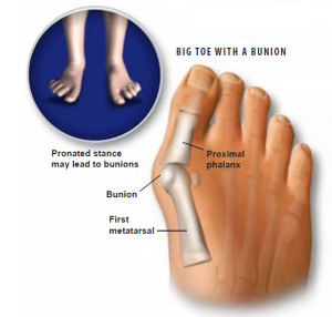
- A bunion (also known as hallux valgus) occurs when the farthest part of the big toe is being pulled inward, towards the other toes. This causes the joint at the base of the big toe to be pushed outwards, away from the other toes. This bone experiences a hardening as well as an increase in size. This can be extremely painful and can be removed surgically.
- Bunions can be a result of narrow shoes, arthritis, and genetic factors.
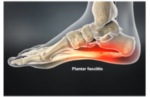
Plantar Fasciitis
- Plantar fasciitis occurs when the tissue supporting the bottom of the foot (the plantar fascia) is strained or injured. The plantar fascia runs right from the heel to the toes. Repeated stress to this tissue can cause tiny tears, resulting in swelling and pain.
- Plantar fasciitis normally presents with pain on the heel or the bottom of the foot.
Flat Foot
- Flat foot is a result of fallen arches in the foot. The arch in the foot is on the inside portion of the foot where it curves up slightly. This arch is made up of tendons that give support to the foot. With flat feet the arches have fallen, therefore no distinct curve is seen on the foot. The tendons are not working correctly to hold the arch up or there is too much weight bearing down on the tendons.
Knee Joint
Anterior Cruciate Ligament
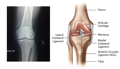
- The ACL (anterior cruciate ligament) is a commonly injured ligament in the knee. The ACL connects the bottom of the femur (thigh bone) to the top of the tibia (shin bone) within the knee joint. The ACL runs on an angle from the top back part of the knee to the lower front part of the knee, within the joint.
- The ACL works to keep the tibia (shin bone) from sliding forward on the femur (thigh bone). If this motion happens, what is called “hyperextension” occurs. When this happens many people say that their knee “bent backwards”, actually going past a straight position and bending slightly in the opposite direction that it normally would.
Posterior Cruciate Ligament
- The PCL (posterior cruciate ligament) is the sister ligament to the ACL, also connecting the bottom of the femur (thigh bone) to the top of the tibia (shin bone) in the knee joint. The difference between the two ligaments is that the PCL runs on an angle from the bottom back of the knee to the top front of the knee (opposite to the ACL). The PCL is injured less often than the ACL.
- The PCL works to keep the tibia (shin bone) from sliding backward on the femur (thigh bone).
Medial Collateral Ligament
- The MCL (medial collateral ligament) runs vertically along the inner leg at the knee joint. The MCL connects the inner portion of the bottom of the femur (thigh bone) to the inner portion of the top of the tibia (shin bone).
- The MCL gives side-to-side stability for the knee and works to limit the knee being knocked sideways towards the other leg (a position similar to knocked-knees).
Lateral Collateral Ligament
- The LCL (lateral collateral ligament) is similar to the MCL but on the outer part of the leg at the knee joint. The LCL connects the outer portion of the bottom of the femur (thigh bone) to the outer portion of the top of the tibia (shin bone).
- The LCL gives side-to-side stability for the knee and works to limit the knee being knocked outwards, away from the other leg (a position similar to bowlegged).
Meniscus
- Within the knee there are two structures called the medial meniscus and the lateral meniscus. These structures are found in between the femur (thigh bone) and the tibia (shin bone). The medial meniscus is on the inner half of the knee, while the lateral meniscus is on the outer half of the knee. The menisci act almost as pillows to absorb shock and protect the bones from rubbing against each other. The medial meniscus is the more commonly injured of the two.
- Damage to the menisci can result in osteoarthritis of the knee.
Patellofemoral Syndrome
- Patellofemoral Syndrome (also known as Patellofemoral Pain Syndrome) is a blanket term to encompass pain caused by the kneecap (patella). The patella is a bone that floats within the hamstring (thigh) muscles and sits within a groove formed by the femur (thigh bone). The patella is supposed to glide effortlessly within this groove, hardly ever deviating from it’s course. With patellofemoral syndrome the patella is not gliding (or tracking) in the correct location on the femur. This causes uncomfortable rubbing on bone, and ultimately the pain that the patient feels. The cartilage between the patella and the femur can also be worn away, creating more friction between the two bones.
- In most cases the patella will be pulled towards the outside portion of the patients leg. One common reason behind this is that the thigh muscles on the outside of the leg are usually much stronger than the thigh muscles on the inside of the leg, which creates an imbalance in the pull of the patella.
Osteoarthritis of the Knee Joint
- Osteoarthritis at the knee is an inflammation of the knee joint as a result of bone on bone rubbing. The bones that rub together are the femur (thigh bone) and tibia (shin bone). On the ends of each of these bones there should be cartilage as a protective barrier to create a smooth surface for the bones to glide on, but with osteoarthritis this cartilage has worn away.
- The majority of patients present with osteoarthritis on the medial (or inside) portion of the knee.
Shoulder Joint
Shoulder Dislocation/Subluxation
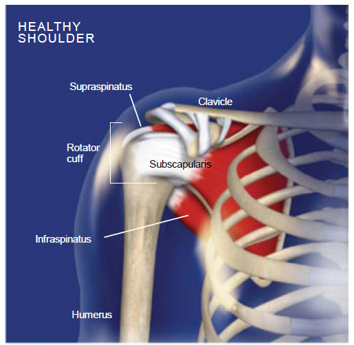
- The shoulder joint is made up of three bones: the scapula, the humerus and the clavicle. The humerus is the bone that forms the upper arm, and the scapula is commonly called the shoulder blade. The scapula has a shallow groove called the glenoid fossa where the head (or end) of the humerus sits. The head of the humerus is shaped like a ball, so that it can spin and glide easily. This gives us much more freedom and mobility with our arms. However, because the glenoid fossa is so shallow it does not always hold the head of the humerus in place, causing the humerus to pop over the edge of the groove. This is a dislocation.
- A shoulder subluxation is when the head of the humerus pops off of the glenoid fossa briefly and then pops right back into place. This can damage tissues by stretching or tearing them.
- Dislocations can weaken the tissues of the shoulder joint by tearing them and stretching them out of place.
Frozen Shoulder
- Frozen shoulder (also known as adhesive capsulitits) is the stiffening of tissues around the shoulder joint. Frozen shoulder can be a result of injury, surgery, or general wear and tear. This condition usually has a steady onset, but takes a long period of time to heal. Frozen shoulder always involves a decreased range of motion for the shoulder joint, not allowing the person to move their shoulder in ways they normally would. This decreased range of motion is a result of stiff tissues and can also involve the buildup of scar tissue in the joint.
Elbow Joint
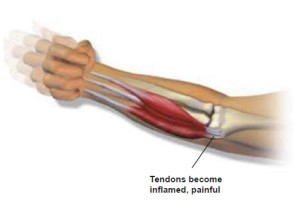
Tennis Elbow
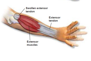
- Tennis elbow (also known as lateral epicondylitis) is an injury that occurs on the outside portion of the elbow. Tennis elbow is very similar to golfers elbow, but occurs on the other side of the elbow. This condition occurs when there is an injury to the muscle or tendon that attaches to the elbow, or through overuse. The injury to the tissue causes inflammation and pain. This condition can occur all of a sudden or develop over a longer period of time.
Golfers Elbow
- Golfers elbow (also known as medial epicondylitis) is an injury that occurs on the inside portion of the elbow. Golfers elbow is very similar to tennis elbow, but occurs on the other side of the elbow. This condition occurs when there is an injury to the muscle or tendon that attaches to the elbow, or through overuse. The injury to the tissue causes inflammation and pain. This condition can occur all of a sudden or develop over a longer period of time.